Friday, April 18, 2014

Ways Laughter And A Confident Smile Are Life Changing

"You don't stop laughing because you grow old. You grow old because you stop laughing."


BECAUSE IT USUALLY HAPPENS SPONTANEOUSLY, we don’t often think about smiling or laughing. However, they both really do make us feel better—and, studies continue to reveal more of their many benefits.


This picture shows a close up view of an 8 ball used for playing pool.It boosts your immune system: 

Smiling really can improve your physical health, too. Your body is more relaxed when you smile, which contributes to good health and a stronger immune system.

This picture shows a blue number 2 inside a hexagon. Smiles are contagious:

It’s not just a saying: smiling really is contagious, scientists say. In a study conducted in Sweden, people had difficulty frowning when they looked at other subjects who were smiling, and their muscles twitched into smiles all on their own.

This picture shows a black number 3 inside a circle.  Smiles relieve stress:

Your body immediately releases endorphins when you smile, even when you force it. This sudden change in mood will help you feel better and release stress.


This picture shows a white number 4 inside a rectangle. Forcing yourself to smile boosts your mood:

Psychologists have found that even if you’re in bad mood, you can instantly lift your spirits by forcing yourself to smile.

This picture shows a black number 5 inside a circle. It's easier to smile than to frown:

Scientists have discovered that your body has to work harder and use more muscles to frown than it does to smile.

This picture shows a white number 6 inside a black circle. Smiling lifts your spirit and those around you:

Life is full of ups and downs. Smiling can reduce distressing emotions and be an invaluable tool in moving forward with our heads held high. And equally important, grins and chuckles can calm and comfort those around us. Here’s an example of how someone else’s smile (laugh) can lift YOUR day… I dare you to watch this video without smiling:


This picture shows a black number 7 inside a badge symbol. Smiles are exercises for  your facial muscles:

53 muscles are used when you smile. Other smiles can take up to 5 muscles. So start working those muscles more often!

This picture shows a white number 8 inside a rectangle. Babies are born with the ability to smile:

Babies learn a lot of behaviors and sounds from watching the people around them, but scientists believe that all babies are born with the ability, since even blind babies smile.

This picture shows a black number 9 inside a circle. Smiling helps you get promoted:

Smiles make a person seem more attractive, sociable and confident, and people who smile more are more likely to get a promotion.

This picture shows a black number 10 inside a circle. Smiling fights stress:

If you're feeling burnt out and tired of the routine, just smile. When you smile, your body releases endorphins (aka “Happy Hormones”), even if it’s a fake smile, this sudden mood change will relieve stress.

This picture shows the number 11 inside a circle. Smiling makes you look really pretty:

Who needs makeup when you have this secret ingredient to instant glam? A research study by Orbit Complete revealed that 69% of people find women who smile more attractive than women who wear a lot of makeup.


This picture shows the number 12 written in white inside a square. Smiling may help you look younger:

That elusive fountain of youth may not be found in surgeries or potions—rather, a study suggests that the path to looking younger may be found through our smiles.


Mark Twain once wrote, “Wrinkles should merely indicate where smiles have been!” The act of smiling is a very uncomplicated and important human activity. It really isn’t important that your smile be perfect- but that you feel good about it and you give it away! If your stingy with your smile because it’s something less than what you want it to be, I can help – Smiles are the best that you can put on!


Sunday, December 29, 2013

THE VICIOUS CYCLE OF TEMPORO-MANDIBULAR-JOINT DYSFUNCTION

The Vicious Cycle of TMJD, 

And How It Affects Its Treatment 


Ramin Mehregan GNM Dentist, headaches, TMJ pain, neck and shoulder pain,

Temporomandibular Joint Dysfunction (TMJD) arises when the harmony of teeth, facial muscles, and the temporomandibular joints no longer exist. Some of the clinical symptoms are:

  • Headaches
  • Neck and shoulder aches
  • Ringing in the ears and ear congestion
  • Clenching/grinding
  • Limited opening/movement of the lower jaw
  • Dizziness/vertigo
  • Clicking/popping of the jaw joints
Patients suffering from these symptoms have learned to accommodate and to live with the daily pain. They have become "the chronic wanderers" going from one medical doctor to the other in the hopes to find an end to their daily misery. Regrettably when competent clinicians cannot find an organic basis for these symptoms, they often suggest it might be psychogenic and/or readily choose the pharmacological route to appease their patients. As these patients are told and retold that there seems to be no tangible basis for their pain, their anxiety, anguish and uncertainty mounts sometimes to levels where depression sets in and they turn to drugs and alcohol to alleviate their feeling of despondency and their symptoms. This in turn can lead to disruptions in work place, relationships and personal achievements. In extreme cases and rare occasions even contemplation of suicide.


The Vicious Cycle


TMJD is a chronic degenerative disease which often takes years to develop. There are many contributing factors that if not diagnosed can and will affect the outcome of its treatment. Every person/patient has a unique personality and biological make-up, and it must be considered when discussing cause and treatment of TMJD. 

The three factors affecting treatment and consequently the outcome are:
  1. Structural
  2. Emotional
  3. Biochemical
A person suffering from TMJD is affected by these three factors in some shape and form and seldom to an equal extent. Hence, it is fundamental when diagnosing TMJD, to asses the relative impact of each of these factors. Many patients may remain oblivious of any symptoms until one day they wake up with, for example an excruciating headache. It is like filling a cup of water to the rim and one keeps adding droplets until all of the sudden there is a spill over. Similarly, many different lifestyles can combine which could aggravate TMJD - such as structural imbalance, stress, depression, fatigue, nutrition and even simply routine check up visits to the dentist. Any of these are like "the single drop" in the cup of water. 

Structural


One of the primary root component that is always present in TMJD is structural imbalance. Structural Imbalance. We often hear this phrase used along with the diagnosis of our aches and pains. But what does it mean? The best way to explain it is to start from the opposite end, "Structural Balance". 
Structural Balance refers to the optimal positioning of our bones and joints. This means when weight or pressure is applied to a part of the body, it is effectively passed through the structures, e.g. bones, joints and muscles. These structures are designed to hold weight well in a particular direction and not so great in other directions. Structural imbalance is simply when bones, joints and muscles are not in their optimal position when weight and pressure is applied. 
  
The structural imbalance in the jaw is also known as "mal-occlusion" or simply a "bad bite". This means that the upper and lower teeth do not close together in the right way. Just as a chair needs four legs in order for one to sit on without falling over, similarly all teeth need to fit firmly together to support the muscles in the face and the muscles used for chewing and swallowing. Teeth are an extension of the skeletal system. The lower jaw is related to the skull via the Temporomandibular Joints (TMJ) and held in place by a collection of muscles. Both the position of the TMJoints and the muscles are determined by the way the upper teeth and lower teeth fir together. When teeth are together they are in occlusion. The occlusion dictates the relation of the jaw to the skull which is also know as the cranio-mandibular relationship.
In the presence of mal-occlusion the skeletal system is in disharmony with neuromuscular system. This means that the teeth/occlusion is forcing the TMJoint's and muscles into a strained position. The aforementioned state increases the natural demand to the muscle and nervous system beyond their natural biological capacity and places them in a spasm cycle.
It is just like walking with a pebble inside your shoe. After a few yards the pain becomes annoying and will twist and torque your hip into all kind of different positions to avoid walking on the pebble inside your shoe (this manipulation is known as accommodation/avoidance). After few more painful steps the pebble will start to feel like a large stone and your foot, leg and body is left in tremendous agony and distress. The former metaphor can be related to what happens to the muscles of the face and jaw (oro-facial muscles) when they are forced into an unnatural position by virtue of a "bad bite". Eventually the muscles become strained and painful. This strain on the muscles causes an alteration in their shape and tonus. To compensate for this the other muscles such as the neck, shoulder and back have to accommodate for this unnatural position, which in turn places them into a strain and spasm.

"Muscles under constant strain are in pain"

Emotional


Many medical/dental professionals are still of the opinion that TMJD is a psyhosomatic (psych=mind, soma=body) illness, starting in the mind and manifesting itself in the body. Sometimes humans continually complain of a discomfort or a pain for which a health professional cannot find an organic cause. The pain or the discomfort, however, is very real to the affected person. Physical complaints with no apparent medical basis may be a reflection of a stress in social settings, relationships, work environment and or other stressful situations for example. Stress, as it affects the body and the mind, has an effect on some illnesses, such high blood pressure, ulcers, and can influence as how humans perceive the symptoms of the illness, how he or she deals with the illness, and the rate and success of recovery.
It is believed, however, that for most part TMJD has its origin from a structural imbalance or "somatopsychic". The resulting pain in the muscles (soma) causes a feeling of anxiety, tension and stress (psyche). There have been numerous studies on how the impact of being sick affects us mentally and emotionally. For instance emotional reactions when having a daily headache could include:

  • Inability to perform up to ones personal and public expectations
  • Anxiety
  • Depression
  • Frustration
  • Lack of energy
  • Anger and hyper-irritability
  • Fear of the unknown
Thus, it is apparent how coping with a chronically painful condition such as TMJD can place someone under tremendous amount of emotional stress. Mind and body is a complex interconnected highway, so that even though the problem may stem from a structural imbalance in the jaw, the resulting discomfort will affect the mind as well.

"My silence is just another word for My pain"

Biochemical


The human body and mind has certain arsenal to its possession when in pain and in stress. In pursuance of pain and stress it prepares itself for either "fight or flight". These are short term positive coping mechanism in the presence of stress. Unfortunately when in pain, under stress and emotional constraints at the same time, we tend to add to the problems by adopting less favorable habits such as:
  • Over-eating
  • Drug and alcohol consumption
  • Medications - prescribed and non prescribed
  • Sleeplessness
  • Not exercising
Soon the body adapts itself to long term chronic stress. In this second stage, the body's defense hormones operate in an above-normal rate. In addition to the aforementioned ill-habits, just like your car would eventually run out of fuel, the body will soon be depleted from its natural fuel supply. At the third stage, exhaustion sets in and the body breaks down. This stage makes it very difficult to treat a chronic disease such as TMJD. The astute clinician treating TMJD may be successful in properly aligning the structural imbalances but some deep seated emotional and bio-chemical disorders need to be managed by appropriate experts or the treatment will fail.


With TMJD the body is placed in constant stress. The extent and severity differs from individual to individual. Some people may have an occasional click in their TMJoints and some some may suffer from constant, severe, daily pain. In either case, the body prepares itself to fight against the stressor. Biochemical changes occur to provide the body with energy to compensate and adapt. This energy is limited however, and eventually will be depleted at a faster rate, especially in the presence of ill-habits and some emotional conditions. This in turn will affect the success of treatment and its longevity. It is of utmost importance that the treating clinician has the ability to recognize the biochemical and emotional factors of their patient and through discovery and co-communication plan for a successful treatment.

"It is important to know the person who has the disease as to know the disease the person has." 


Monday, November 18, 2013

THE 40 POUND HEAD - DAMAGING EFFECTS OF FORWARD HEAD POSTURE

Forward head posture and its effects on health and the craniomandibular complex

Ramin Mehregan GNM dentist, headaches, TMJ pain, neck and shoulder pain

The effect of posture on health is becoming more evident. “Spinal pain, headache, cranio-mandibular-joint effects, mood, blood pressure, pulse and lung capacity are among the functions most easily influenced by posture. 
One of the most common postural problems is the forward head posture (FHP). Since we live in a forward facing world, the repetitive use of computers, TV, video games, trauma, compromised occlusal plane and even backpacks have forced the body to adapt to a forward head posture. It is the repetition of forward head movements combined with poor ergonomic postures and/or trauma that causes the body to adapt to forward head posture.

A review of literature substantiates that "For every inch of forward head posture, it can increase the weight of the head by and additional 10 pounds." It's not uncommon to have TMD patients walk into my office supporting a 10-12 lb head that has migrated 3 inches forward of their shoulders. It isn't difficult to recognize prior to any palpation that their cervical muscles are in a losing battle attempting to isometrically restrain 40-42 pounds against the unrelenting forces of gravity.


Ideally, the head should sit directly on the neck and shoulders, like a golf ball sits on a tee. The 
weight of the head is more like a bowling ball than a golf ball, so holding it forward, out of alignment, puts a strain on your neck and upper back muscles. The result can be muscle fatigue and all to often an aching neck. Head forward posture can add up to thirty pounds of abnormal leverage on the cervical spine. This can pull the entire spine out of alignment. 


Because the neck and shoulders have to carry this weight all day is an isometric contraction, this causes neck muscles to loose blood , get damaged, fatigue, strain, cause pain, burning and fibromyalgia. When spinal tissues are subject to a significant load for a sustained period of time,
they deform and undergo remodeling changes that could become permanent.


It also has been noted that forward head posture
may result in the loss of 30% of vital lung capacity.
These breath related effects are primarily due to the
loss of the cervical lordosis which blocks the action of the 
hyoid muscles, especially the inferior hyoid responsible for helping the first rib during inhalation. Proper rib lifting action by the hyoids and anterior scalenes is essential for complete aeration of the lungs.


Head Posture and Cranio-mandibular posture

The relationship of the mandible to the cranio-maxillary complex, the temporomandibular joints, the atlas and the cervical and thoracic vertebrae are orthopedic in nature. In addition the shoulders, clavicles and sternum are all affected during the action of mastication and swallowing. Many of theses structure either share the same neuro-muscular system or have close commonalities. 
Patients with deep bites, retruded lower jaws (Class II mal-occlusion), deficient vertical dimension, narrow introral arches typically present with a forward head posture and a variety of symptoms related to TMJD. 

Often seen as a structurally subtle body segment, the neck is burdened with challenging task of supporting and moving the human head. Because of tension and poor postural relationship of the craniomandibular complex and habits inherent in today's workplace and society it comes as no surprise that associated neuromuscular disorders rank high as the most common pain generators. Correction of the upper cervical area and the mandibular relation to the cranio-maxillary complex is key to stoping and reversing degenerative joint disease and pain from headaches, breathing abnormalities, TMJ dysfunction and other postural effects. Any loss of function sets off reactions within the body's open, dynamic system which manifests as structural abnormalities throughout the entire body.


Wednesday, November 6, 2013

TEMPORO-MANDIBULAR-JOINT DYSFUNCTION AWARENESS

TMJ Dysfunction Awareness


In 1997 November was officially declared as the "Jaw Joints-TMJ Awareness month in the United States of America.
Temporomandibular Joint Disorders, commonly referred to as “TMJ,” afflict millions of people not only Americans but worldwide, both children and adults of both sexes and all races. TMJ dysfunction is a painful and often disabling disorder that emanates from the Jaw Joints and can affect the health of the entire neuro­musculo-skeletal system often spreading and dysfunction throughout the entire body. 

Understanding the complex relationship of temporomandibular disorders to overall health requires broad understanding of not only the anatomy and physiology of the head and neck but also the cervical spine and upper quarter complex.


There are over 120 known signs and symptoms related to TMJ dysfunction. Some of the extra oral signs and symptoms include:


  • Jaw joint noises-clicking, popping, grading noises
  • Headaches
  • Sore muscles of the face and jaw
  • Limited ability to open the mouth
  • Teeth sensitivity to temperature
  • Ear symptoms such as fullness, ringing in the ears
  • Dizziness
  • Facial asymmetry
  • Pain behind the eyes
  • Numbness and tingling of the hands and fingers
  • Neck and shoulder pain
  • Lower back pain
  • Postural issues
  • Nervousness
  • Insomnia

Some of the intra oral signs and symptoms may include


  • Crowded and crooked teeth
  • Narrow upper and lower arches
  • Worn lower front teeth
  • Deep bite (over lapping of upper front teeth over lower front teeth)
  • Cross bites
  • Anterior open bites
  • Missing teeth
  • Broken back teeth
  • Tongue thrust habits
  • Grinding and clenching
  • Phantom tooth ache

The aforementioned are just a small example of the myriad signs and symptoms related to TMJ dysfunction. If you are experiencing any of these symptoms and non of the medical interventions have helped or you are a victim of over prescription of an array of medications, you are not alone. According to National Institute of Dental and Craniofacial Research NIDCR, TMJ disorders are the second most common pain causing musculo-skeletal conditions after chronic lower back pain. It is estimated that 5-12% of the population is affected by TMJ dysfunction.

This awareness can be increased among all the countries and not just the USA (where this awareness month was instituted), and in particular those in a position to help treat, insure, provide appropriate research, and ultimately to prevent this scourge everywhere.

Monday, September 23, 2013

THE IMPORTANCE OF OBJECTIVE EVALUATION IN THE TREATMENT OF TMJD

The Rationale For The Use Of Bioelectronic Instrumentation In The Treatment Of Temporomandibular Dysfunction (TMD)

Why I use the K7 Evaluation System1 


Initial diagnosis, whether be it in dentistry or medicine, relies  greatly on information gathered from a thorough history and from comprehensive clinical examination. Choosing the correct route of treatment is based on an understanding of the pathogenesis of the disease being treated after a correct diagnosis has been confirmed subjectively and more important objectively. It has been shown repeatedly that diagnosis based on subjective evaluation alone can lead to gross errors in the diagnosis. In almost every discipline of the health care system bioelectronic instrumentation has become a paramount and necessary component to aid in the objective evaluation process. It aids in the progress and confirmation as additional hard data are obtained. This data/information will aid the doctor in arriving at a diagnosis, quantifying the parameters of the illness being treated, determining the right therapy, and evaluating therapeutic outcome. For example a cardiologist may prescribe an EKG under various condition to evaluate the stress that is being excreted on the heart.
In today's modern society it is no longer reasonable, customary, or acceptable for the physician to rely solely on history and subjective evaluation using visual examination and palpation.

Why is then that within the healthcare discipline of dentistry, with a small exception, has remained reserved and reticent to enter the age of electronic measurement/instrumentation?

Why is it that the majority of the dental professionals rely only on minimal dental radiography (if any), visual examination, audible evaluation, hand articualtion of dental models and hand manipulation of the lower jaw into a "centric relation" when it comes to the treatment of Temporomandibular Joint disorders?

Isn't the ultimate therapeutic goal for any treatment regimen to improve health by returning the body to an optimized state of physiologic comfort and function?


The role of Myotronics K7 Evaluation system in the objective diagnosis of TMD

The K7 Evaluation System has the capacity to record, analyze, and interpret gross and fine movements of the mandible and joint sounds, while monitoring the activity of the masticatory muscles in life time. This enables the Gneuromuscular clinician to create a measured occlusal position in the six dimension of mandibular movement with knowledge of the masticatory muscular imlication and disc/condyle relation of that particular occlusion. This philosophy abdicates from the historical philosophies of manual manipulation and hand articulation which obscured the mandibular and muscular functional implications. The use of electronic measurement assure an objective evaluation and diagnosis and assures that the created occlusion is physiologically sound. The K7 Evaluation system provides the clinician with three technologies for measuring, displaying and storing objective data on physiologic and anatomical status and function:

  1. Jaw Tracking
  2. Electromyography
  3. Joint Sonography 

The primary components of TMD and the treatment modalities employed involves mandibular movement, dental occlusion, masticatory muscle and Temporomandibular Joint function. With objective data, the patient's condition before treatment can be assessed, a therapeutic plan created to effect the needed changes and the results of treatment analyzed for efficazy. Electronic instrumentation provides reproducible and quantifiable data needed for treatment.

Jaw Tracking employing the K7 instrumentation

Mandibular tracking allows the clinician to detect and scrutinize minuscule mandibular movements in three dimensions at habitual/pathological centric occlusion and rest and compare this to post TENS (Trans Electrical Neural Stimulation) true physiologic rest position and determined Optimized Occlusal Position2 .
Scans/data are obtained pre-TENS therapy and post-TENS therapy.


Pre-TENS and Post-TENS Scans

Scan 1:                                                                                                                                    
It records the patient's normal opening and closing in the Sagittal and Frontal mode simultaneously. 
It gives an indication of:
  • Normal closure into occlusion
  • Accommodated closure into occlusion
  • Any interferences on the mesial/distal or buccal/lingual facing cusp inclines
  • Precise terminal centric contact
  • Musculo-skeletal strains
  • Maximum full range of opening
Scan 2:                                                                                                                                    
It is designed to record speed (velocity) of mandibular movement in both sagittal and frontal mode during opening and closing. 

It gives an indication of:
  • Functional joint anatomy (condyle, disc, eminence)
  • Correlation between joint and occlusion
  • Clicks exact location (slow down in velocity)
  • Quality of terminal tooth contact
  • Opening symmetry
  • Occlusal stability and interferences
  • Slow movements, jerky/erratic movements
  • Mandibular deviations
  • Muscular imbalances
  • Joint health and condition
Scan 3:                                                                                                                                    
It shows the three dimensional movement of the mandible of vertical, antero-posterior (AP), and lateral movements.

It gives an indication of:
  • Stability of habitual REST
  • Vertical position from habitual centric occlusion (CO)
  • AP deviations and lateral deviations
  • Muscular and joint stability/instability
  • Habitual pathway of closure to terminal tooth contact
  • Suggestive of possible "Clencher profile"
Scan 4 and Scan 5 are combined and taken post-TENS (it will be explained in greater detail subsequently)

Scan 6:                                                                                                                                    
It shows mandibular movement during swallowing and identifies tongue activity.

It gives an indication of:

  • The path of closure to centric occlusion during swallowing
  • The position of the mandible during swallowing movement
  • Position of the tongue during swallowing
  • Lateral and anterior tongue thrust
  • Occlusal stability/instability
Scan 7 resembles Scan 2 and it is taken post-TENS

Scan 8: (is also taken post-TENS and orthosis therapy)                                                           


It shows mandibular movement during functional chewing cycles.

It gives and indication of:

  • Quality of the terminal intercuspal position during function and resting mode
  • Left and right lateral movement symmetry
  • Measures mandibular position within the cuspal inclines
  • Envelop of chewing motion
  • Occlusal guarding
  • Preciseness of centric terminal occlusion
  • Muscle and joint health
Scan 9:                                                                                                                                    
It is an EMG (Electromyograh) display of the muscle activity at postural habitual rest before TENS.

It gives and indication of:

  • Showing relative resting activity/firing of masticatory and cervical muscles
  • Degree of muscle accommodation during habitual REST
  • Muscle hyperactivity due to malocclusion and/or fatigue
  • Postural/cervical misalignments
  • Clencher profiling
  • Temporalis muscle and its relationship to mandibular posture
  • Masseter muscle activity as it relates to tooth contact
  • Cervical muscles as it relates to mandibular and head posture
  • Digastric/suprahyoid muscle and its relationship to arch shape and tongue posture
Scan 10:                                                                                                                                  
It is an EMG display of muscle activity post-TENS.

It gives and indication of:

  • Degree of muscle relaxation at Physiologic REST
  • Muscle rest due to malocclusion and/or rest
  • Postural/cervical misalignments
  • Clencher profiling
  • Muscle recruitment
  • Not all low EMG's are indication of normalcy
Scan 11:                                                                                                                                  
It is designed to record the quality of muscle recruitment of the temporalis and masseter muscles respectively during function (clenching).

It gives an indication of:


  • Quantitative display muscle activity during function
  • Relative amount of work each muscle is capable of excreting
  • Verify effect of orthosis therapy
  • Possible occlusal imbalances
  • Lack of posterior support
  • Fatigued muscles vs. muscle recruitment ability
  • Quality of occlusal proprioception
  • Clencher profiling 
  • Comparison of relative muscle firing between left and right
Scan 12:                                                                                                                                  
It is an electromyographical demonstration of muscle activity displaying mandibular torque. Used in the refinement of occlusal anatomy to assure balanced occlusion.

It gives an indication of:


  • First tooth contact with simultaneous EMG to assist in identifying where to adjust the bite in micron levels
  • Identifying diagnostically initial deflecting contacts (prematurities)
  • Monitors early motor unit recruitment as the patient closes from rest position through freeway space into first tooth contact
  • Allowing easy interpretation of first tooth contact
  • Shows synchronous balanced muscle recruitment during closure
Scan 13:                                                                                                                                 
It is used to record mandibular range of motion including the maximum vertical opening and can be used both pre and post-TENS. 

It gives an indication of:
  •  Level of improvement post-TENS
  • Deviations and asymmetrical movements due to joint and muscle pathology
  • Restrictions during maximum range of mandibular motions
  • The quality of opening and closing in sagittal and frontal paths
Scan 15:                                                                                                                                                                   
It is a combination of jaw tracking and sonography allowing to associate sound patterns with real time opening and closing cycles of the mandible.

It gives and indication of:

  • Alterations in joint tissue morphology
  • Thickening of articular surface
  • Macroscopic remodeling
  • Condylar deviations due to articular disc displacement
  • Level at which clicks occur
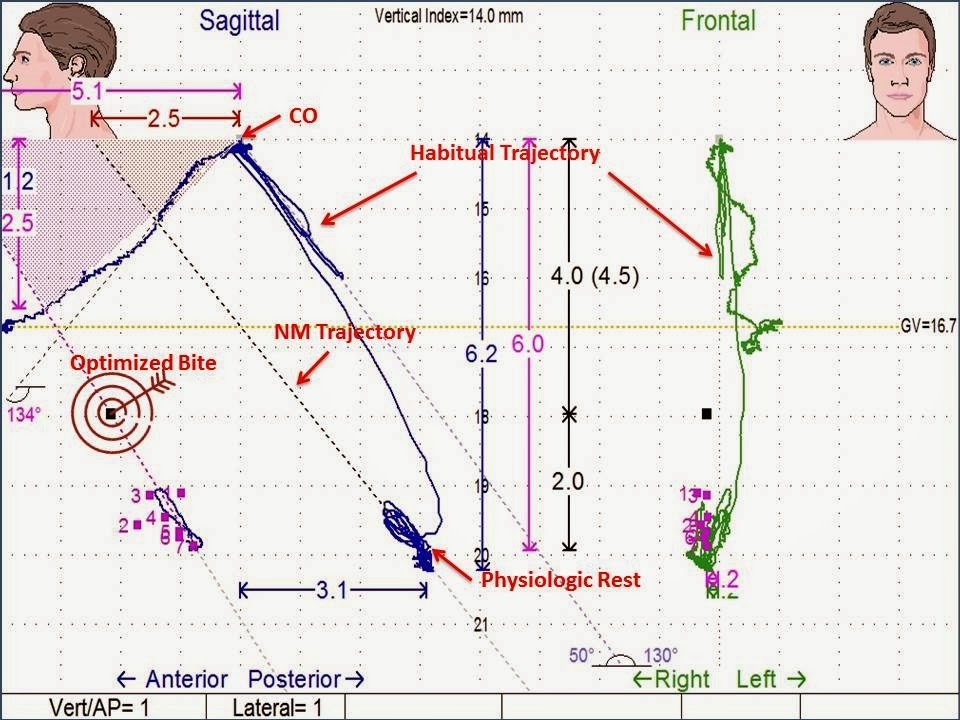
Scan 4/5: And the Optimized Bite3 
It is a combined recording of sagittal, frontal and lateral positioning of the mandible as it relates to the maxilla (ultimately skull) from centric occlusion over time. Scan 4/5 is the defining scan that distinguishes a NM dentist from other clinical dentists.


It gives indication of:


  • The quality of physiologic rest after TENS
  • Location habitual trajectory, the classic NM trajectory, and the Optimized trajectory
  • Quality of vertical and AP pulse
  • Mandibular shifts during bite recording
  • Disc recapturing
  • EMG recordings
  • Level and stability of muscle relaxation of the masticatory and cervical system
  • Repeatable

What is Bite OPTIMIZATION?

"Optimization is a term used to distinguish itself different than the classical neuromuscular jaw positioning. It is a technique and protocol that uses a systematic bite finding protocol using jaw tracking instrumentation (Myotronics K7,Scan 4/5) to “Optimize the Bite”. It is a bite recording protocol that goes beyond classical TENS bite,modified TENS bite techniques,classic scan 4/5 bite protocols and “modified Scan 4/5 bite”taking protocols developed and pioneered by the originator Dr. Clayton Chan. The Optimized Bite technique and protocol is scientific and can be measured to within 0.1-0.3 mm accuracy."

Conclusion

Bioelectronic measurement instrumentation provides a road map to healthy dental occlusion, neuromuscular system and posture. It provides accurate and objective data of the mandibular and masticatory muscle function during pathology and health providing essential information for proper treatment. However, that said the clinician must develop a protocol in which each specific test adds or refines the treatment. Further the instrumentation only serves as a guide to aid in the diagnosis and treatment protocol, it is the astute clinicians knowledge to verify the data and to establish a proper outcome.

References:

1) K7 Evaluation System, Myotronics Inc. Kent, WA. USA
2) Optimized Occlusion Position, "Clinical and Scientific Validation for Optimizing the Neuromuscular Trajectory using the Chan Protocol", Clayton A. Chan D.D.S., ICCMO Anthology Vol. VII, 2005
3) Optimized Bite Protocol, "NM Bite Refinement, Level 5 K7 Practicum", Occlusion Connections, Las Vegas, NV.


Thursday, August 22, 2013

TMD UNDERSTANDING THE GREAT IMPOSTER

Temporomandibular Joint Dysfunction

Understanding the Great Imposter


Relationship between occlusal proprioception and the neurologic effect

Understanding the relationship between occlusal proprioception and the neurologic effect at higher levels is essential in the understanding of TMJD symptoms that these symptoms are usually not in the joint itself, but of neuromusculature nature. Primarily in the masseter, temporal, medial and lateral pterygoid and digastric muscles. Evidence suggests that the basis of symptomatology is a disturbance of proprioception at a mid brain level.

Proprioception and the Brain Stem Reticular Formation

Located in the central part of the brain stem, the reticular formation is a tiny network of nerves the size of man’s little finger. All the major nerve trunks in the body have tendril like branches to the reticular formation. Afferent (carry nerve impulses from receptors or sense organs towards the central nervous system) sensory signals from all parts of the body go to the cerebral cortex by direct pathways ascended through the brain stem; however, they send collateral nerves (side branches) to the reticular formations. The cerebral cortex is the site of perception, thought and ability to respond to a stimulus with anything more than a simple reflex reaction. But directly stimulating the cortex will not awaken the brain. The reticular formation, also know as the reticular activating center (RAC) simply has one action to arouse the brain.


The Trigeminal Nerve

The Trigeminal Nerve (the fifth cranial nerve) is the largest of the cranial nerve, it contains 60% of the total nerve tissue of the twelve cranial nerves. It gives origin to three major divisions:
  1. The Ophthalmic V1       
  2. The Maxillary V2
  3. The Mandibular V3  
It is the sensory nerve of the face, most of the scalp, the teeth, the mouth, nasal cavity, and the temporomandibular joint and it carries proprioceptive impulses from the masticatory muscles. It's motor branches supply the muscles of mastication and the other muscles such as the mylohyoid, anterior belly of the digastric, tensor veli palatini and tensor tympani. (An understanding of the TVP and tensor tympani you may refer back to my previous Blog TMD and the EAR and EYE Connection). 
There are many parasympathetic and sympathetic nerve fibers from the other cranial nerves that join branches of the trigeminal nerve. Its influence on the central nervous system is accordingly disproportionate to that of the other cranial nerves. The proprioceptive stimulation of occlusion is the dominant sensory input into the trigeminal system.

The trigeminal nerve is uniquely associated with the ascending activating reticular system. The primary sensory trigeminal fibers terminate in the reticular formation just medial to the spinal trigeminal nucleus. The spinal afferents from all levels terminate in the spinal and sensory nuclei of the trigeminal nerve. In voluntary movements the sensory nerves conduct impulses from the muscle spindle to a sensory area in the brain. Motor nerves then conduct impulses from the motor area to the masticatory muscles. Both nerve systems branch into the reticular activating system. RAS sends down efferent, (carry nerve impulses away from the central nervous system to effectors such as muscles or glands), impulses that either facilitate or inhibit the response. The reflex movement sensory impulses are transmitted immediately to motor nerves in spinal cord. One nerve activates the muscle and maintains its tone and the other nerve sensitizes the muscle spindle. Both voluntary and reflex mechanisms are under reticular activating system control. 


All evidence suggests that the reticular activating system has a most important role in regulating all motor activities in the body. It can modify voluntary muscle movements (controlled by the brain) or the reflex movements (controlled by the spinal cord). The importance of this voluntary and reflex arc on muscles of the face and jaw is seen in TMJD patients.

The fact that the RAS can act on the spinal cord reflexes distinguishes even further the role of noxious proprioceptive occlusal contacts (such as grinding or interferences due to mal-occlusion) as pathogenic sources to muscles. The reflex apparatus has two functions:

  1. First, it generates automatic muscle movements. When noxious stimuli (i.e. occlusal prematurities) arrive at the spinal cord, they are instantaneously passed on to an adjacent motor nerve and travel back to the affected part of the body to jerk it away from the noxious stimuli. This nociceptive  (A nociceptor is a sensory receptor that responds to potentially damaging stimuli by sending nerve signals to the spinal cord and brain) reflex is designed to protect the body part from injury. The avoidance of the prematurity may protect the tooth from a noxious contact but significant accommodation of muscles and joints is often required.
  2. The second function of the reflex system is to keep the muscles ready for action by maintaining muscle “tone”. This muscle tone postures the body part close to the area where function will occur. The muscle is in a state of partial contraction in anticipation of the work to be done. The muscle spindle regulates the resting tone. When muscle contracts, it squeezes the spindle; when the muscle relaxes the pressure on the spindle loosens. Change from normal tone causes the spindle to send signals via the sensory nerve to the spinal cord. The signal then excites a motor nerve to correct the contraction or relaxation of the muscle. This feedback system automatically maintains proper muscle tone.
    The tone of the muscle is adjusted to the functional demands on the muscle by nerve impulses which regulate the sensitivity of the spindle. Tactile sensibility for the trigeminal nerve is mediated by the sensory nucleus. Pain and thermal sensibility is mediated by the spinal nucleus or the trigeminal nerve. Under normal circumstances the reticular formation exerts a restraining influence on impulses conducted by the trigeminal as well as by the spinal nerves. It is obvious that if the tonic activity of the reticular formation is disturbed by noxious proprioceptive interferences the restraining influence of the reticular activating system on sensory impulses conducted by the trigeminal nerve will be significantly affected.
It is important to have a thorough understanding of the afferent and efferent neuromuscular connection between occlusal proprioception, the trigeminal nerve, reticular activating formation, cerebral cortex and skeletal muscles. Many of the dysfunctional conditions suffered by patients with pathologic occlusion is explainable when the neuromuscular mechanism is understood.


References:
1.  Chan C.A.: "Diagnostic Principles" Level 2 Occlusion Connections, Las Vegas, NV.
2.  Jankelson, R., "Neuromuscular Dental Diagnosis and Treatment", published by Ishiyaku EuroAmerica, ST Louis, MI. 2nd edition 2005